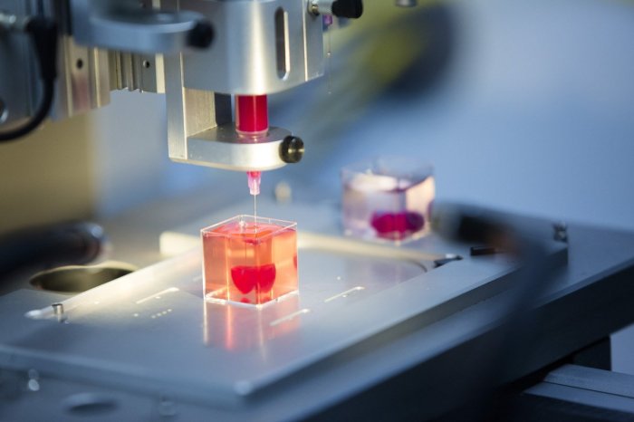
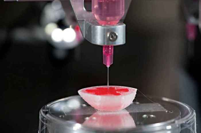
3D printer generates synthetic tissue – it sounds like science fiction, right? But this isn’t some futuristic fantasy; it’s happening now. This groundbreaking technology is revolutionizing regenerative medicine, offering the potential to create replacement tissues and organs for those in need. From bioinks and intricate printing techniques to the ethical considerations of growing human tissue, the world of 3D bioprinting is complex and fascinating. Let’s dive into the details.
Imagine a future where damaged organs can be replaced with perfectly matched, bioprinted tissues. This isn’t just a pipe dream; 3D bioprinting is rapidly advancing, promising to revolutionize healthcare and offer solutions to previously incurable conditions. This technology uses specialized “bioinks” – essentially living cells suspended in a gel-like substance – to build tissues layer by layer. Different printing methods, each with its own advantages and disadvantages, are constantly being refined to create increasingly complex and functional tissues.
Applications of 3D Printed Synthetic Tissues
The ability to 3D print synthetic tissues represents a paradigm shift in regenerative medicine, offering unprecedented opportunities to repair or replace damaged organs and tissues. This technology moves beyond simple tissue grafts, enabling the creation of complex, functional structures tailored to individual patient needs. The implications are vast, spanning numerous medical fields and promising revolutionary treatments for previously intractable conditions.
3D bioprinting allows for the precise placement of cells, growth factors, and biomaterials within a scaffold, mimicking the natural architecture of tissues. This level of control offers significant advantages over traditional transplantation methods, potentially leading to improved integration, reduced rejection rates, and enhanced functional outcomes.
Pre-clinical and Clinical Trial Examples
Several pre-clinical and clinical trials have demonstrated the potential of 3D bioprinted tissues. For instance, researchers have successfully bioprinted functional skin grafts for burn victims, showing promising results in wound healing and scar reduction. In another example, pre-clinical studies have explored the creation of 3D-printed cartilage, demonstrating the potential to repair damaged joints. While still in early stages, clinical trials involving bioprinted tissues for cardiovascular applications are also underway, focusing on the creation of blood vessels and heart patches. These early successes highlight the rapid advancement of this technology and its translational potential.
Ethical Considerations, 3d printer generates synthetic tissue
The use of 3D printed synthetic tissues in human therapies raises several ethical considerations. One key concern revolves around the source of cells used in bioprinting. The use of autologous cells (from the patient themselves) minimizes the risk of rejection, but obtaining sufficient quantities of cells can be challenging. Allogeneic cells (from a donor) offer an alternative but raise ethical issues surrounding donor consent and the potential for disease transmission. Additionally, equitable access to this potentially life-saving technology needs careful consideration to prevent widening existing healthcare disparities. Finally, the long-term effects of 3D printed tissues on the human body are still under investigation, requiring robust and long-term follow-up studies to assess potential risks.
3D Printed Tissues vs. Traditional Transplantation
Compared to traditional transplantation methods, 3D bioprinting offers several advantages. It allows for the creation of tissues with precise architectures and tailored properties, potentially leading to improved integration and reduced rejection rates. The process also allows for the incorporation of growth factors and other bioactive molecules to promote tissue regeneration. Furthermore, it offers the possibility of creating tissues “on demand,” reducing reliance on donor availability and minimizing waiting times for transplants. However, 3D bioprinting is currently a more complex and expensive process than traditional transplantation. The technology is also still under development, and the long-term efficacy and safety of bioprinted tissues need further investigation.
Potential Future Applications of 3D Printed Synthetic Tissues
The future applications of 3D printed synthetic tissues are incredibly promising and extend far beyond current capabilities. The technology holds the potential to revolutionize numerous medical fields.
- Oncology: Creating personalized tumor models for drug testing and developing bioprinted scaffolds for cancer immunotherapy.
- Orthopedics: Producing customized bone and cartilage implants for joint repair and fracture healing.
- Cardiology: Generating functional heart patches and blood vessels for cardiovascular disease treatment.
- Neurology: Developing bioprinted neural tissues for spinal cord injury repair and neurodegenerative disease treatment.
- Dentistry: Creating personalized dental implants and reconstructing damaged jawbones.
- Ophthalmology: Bioprinting corneal tissues for vision restoration.
- Urology: Developing bioprinted tissues for bladder and kidney repair.
Illustrative Examples: 3d Printer Generates Synthetic Tissue
The field of 3D bioprinting has yielded impressive results in creating functional synthetic tissues. While challenges remain, several examples showcase the technology’s potential to revolutionize regenerative medicine and tissue engineering. Below are two detailed examples illustrating the diverse applications and capabilities of this exciting technology.
3D Bioprinted Cartilage
One successful example of 3D bioprinted synthetic tissue is the creation of cartilage. Researchers have successfully bioprinted cartilage constructs using a bioink composed of chondrocytes (cartilage cells) embedded in a hydrogel matrix, often alginate or hyaluronic acid. These hydrogels provide a supportive environment for cell growth and differentiation. The printing process typically involves extrusion-based bioprinting, where the bioink is precisely deposited layer by layer to create the desired cartilage structure. The resulting tissue exhibits a highly organized structure, mimicking the natural extracellular matrix of native cartilage.
The printed cartilage appears macroscopically as a smooth, translucent, pale-yellow structure, its shape determined by the printing parameters and the design file. Microscopically, the tissue shows a well-defined distribution of chondrocytes within the hydrogel matrix. The cells are viable and maintain their chondrocytic phenotype, producing extracellular matrix components such as collagen type II, characteristic of healthy cartilage tissue. The overall architecture is highly organized, with minimal cell aggregation or necrosis. This closely resembles the natural structure of cartilage, a crucial factor in its functionality and integration with the host tissue.
3D Bioprinted Vascularized Skin
Another significant achievement in 3D bioprinting is the creation of vascularized skin grafts. This complex tissue requires a sophisticated approach, as it involves multiple cell types and a complex vascular network essential for nutrient and oxygen delivery. Bioinks for vascularized skin often incorporate endothelial cells (lining blood vessels) and fibroblasts (connective tissue cells) within a biocompatible hydrogel, potentially supplemented with growth factors to stimulate angiogenesis (blood vessel formation). The printing process may involve multiple nozzles to co-print different cell types and create a layered structure, mimicking the dermis and epidermis. Advanced techniques, such as perfusion bioprinting, allow for the creation of pre-vascularized constructs.
The resulting bioprinted skin exhibits good mechanical properties, including tensile strength and elasticity, comparable to native skin. Microscopically, the tissue demonstrates a layered structure, with a well-defined epidermis and dermis. Endothelial cells form interconnected vascular channels, ensuring adequate perfusion and cell viability. The integration of the printed skin with host tissue is facilitated by the biocompatibility of the bioink and the presence of growth factors that stimulate tissue regeneration. The appearance is similar to natural skin, with a smooth, slightly textured surface. However, achieving a full pigmentation that perfectly matches the host’s skin tone is still a work in progress. The successful integration with host tissue, as evidenced by the formation of new blood vessels connecting the printed graft and the surrounding tissue, demonstrates the potential for improved wound healing and skin regeneration.
The ability of a 3D printer to generate synthetic tissue marks a pivotal moment in medical innovation. While challenges remain – particularly in scaling up production and ensuring long-term biocompatibility – the potential applications are staggering. From repairing damaged hearts to growing replacement skin, the future of regenerative medicine is being written, one bioprinted layer at a time. The journey is complex, filled with scientific hurdles and ethical considerations, but the potential to transform lives is undeniably exciting.
 Invest Tekno Berita Teknologi Terbaru

